
Advanced 3D Imaging at Valley Oral Surgery in Livermore and Dublin, CA
Valley Oral Surgery uses the CS 9300 3D imaging system to support accurate diagnosis, treatment planning, and surgical safety.
What Is 3D Dental Imaging?
3D dental imaging, also known as cone beam computed tomography (CBCT), is a specialized type of X-ray technology that creates detailed, three-dimensional views of your teeth, jaw, nerves, sinuses, and facial structures.
At Valley Oral Surgery, we use the Carestream CS 9300 system, which provides high-resolution, low-radiation scans. These images help us plan oral and maxillofacial procedures with greater accuracy and safety.
Compared to standard 2D X-rays, 3D imaging offers:
- More precise anatomical visualization
- Improved surgical planning
- Better detection of bone defects, cysts, or impactions
- Faster, more efficient diagnostics
Why We Use the CS 9300 System
The CS 9300 is a next-generation CBCT platform that allows our surgeons to:
- Capture images in seconds with minimal radiation exposure
- Focus scanning on specific regions to reduce overall dose
- Visualize complex structures (nerves, sinuses, joints) in high detail
- Plan surgeries virtually before entering the operating room
This system enhances every stage of your care — from diagnosis to execution — while improving safety and minimizing surprises during surgery.
When 3D Imaging Is Recommended
3D scans are commonly used to support:
- Dental implant planning: Precisely evaluate bone density, nerve location, and implant angles
- Wisdom teeth and impacted tooth evaluations: Determine root positions, impaction angles, and sinus proximity (wisdom teeth removal, impacted canines)
- TMJ disorder diagnosis: Visualize joint structure, disc alignment, and bone degeneration (TMJ disorders)
- Bone graft and sinus lift planning: Accurately assess bone volume and sinus location prior to grafting (sinus lift, ridge augmentation)
- Oral pathology or cyst detection: Evaluate lesions, cysts, or abnormal growths (oral pathology)
- Trauma cases or jaw fractures: Diagnose fracture lines and plan stabilization techniques (Dental trauma)
Advantages of CBCT Imaging
Using the CS 9300 system offers several key advantages:
- Greater diagnostic clarity compared to traditional X-rays
- High precision in implant and surgical planning
- Lower radiation dose than hospital-grade CT scans
- Non-invasive and completed in a matter of seconds
- Enables patient education through clear, visual explanations
Our surgeons use these scans to guide surgery, reduce risks, and improve outcomes — especially in anatomically complex areas.
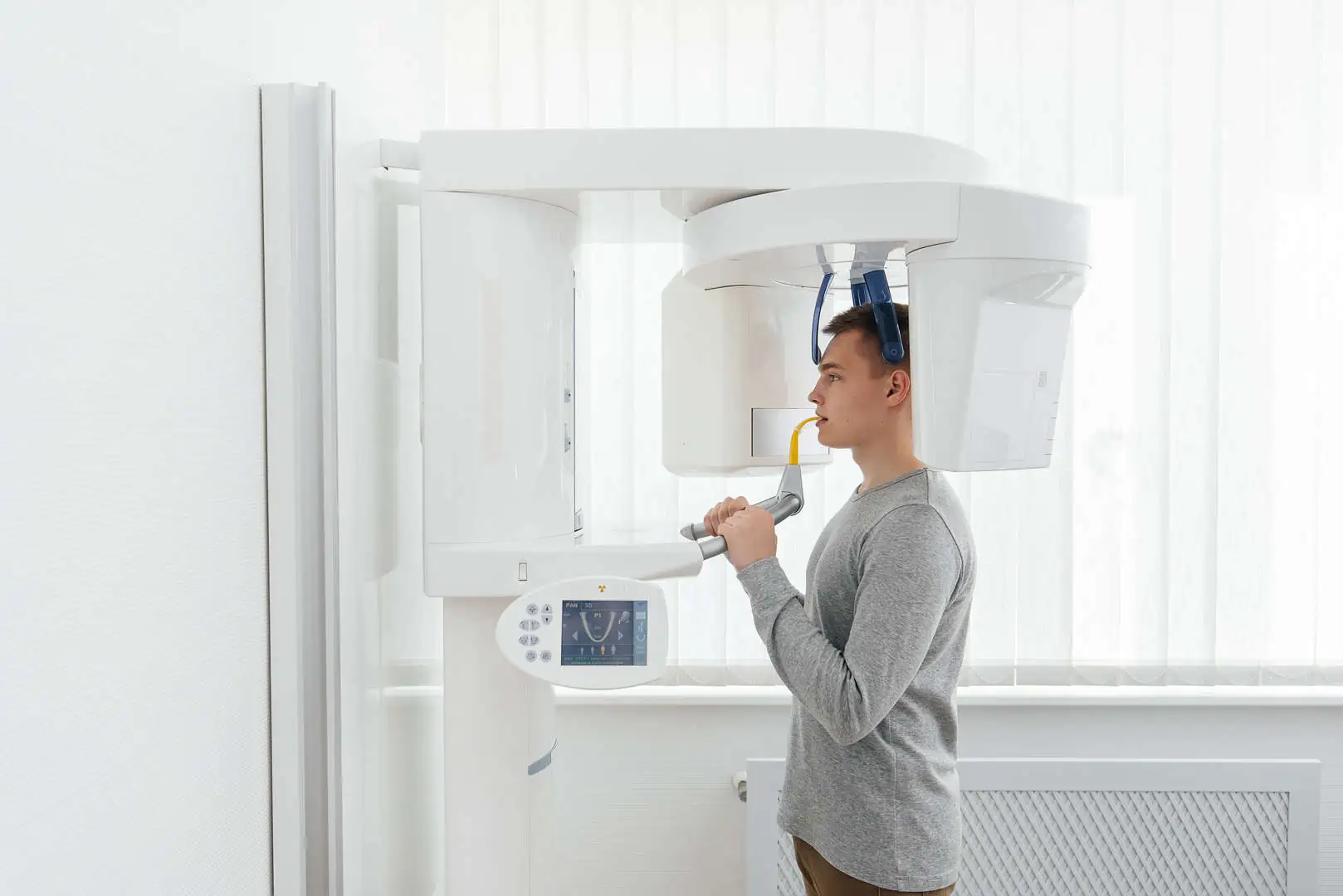
What to Expect During a 3D Scan
A CBCT scan is quick, painless, and requires no special preparation.
- Step 1: You’ll stand or sit in the imaging unit
- Step 2: The scanner rotates around your head for about 10–20 seconds
- Step 3: You won’t feel anything during the scan
- Step 4: Your surgeon will review the images with you on-screen during the same visit
The full imaging and review process is typically completed during your consultation appointment.
Safety and Radiation Considerations
The CS 9300 system is designed to minimize radiation exposure while delivering high-resolution imaging. It uses:
- Targeted scanning areas to avoid unnecessary exposure
- Short scan times for efficiency
- Radiation doses far lower than conventional medical CT scans
For comparison, a CBCT scan delivers about the same radiation as a short airplane flight. It is considered safe and well-tolerated for nearly all patients.
3D Imaging & Diagnostics in Livermore, Dublin & Surrounding Areas
Valley Oral Surgery provides advanced 3D dental imaging at both of our offices in Livermore and Dublin. We also support patients from Pleasanton, Tracy, Castro Valley, Fremont, San Ramon, Hayward, and surrounding Bay Area communities. Our imaging services are available for direct patients as well as referrals from general dentists, orthodontists, prosthodontists, and medical providers.
FAQs: 3D Imaging & CBCT Scans
Is a 3D scan the same as a CT scan?
Cone beam CT (CBCT) is a specialized dental scan that delivers 3D images like a hospital CT, but with a lower radiation dose and a more focused field of view.
How long does the scan take?
Most scans take less than 20 seconds. The entire appointment, including image review, is usually completed within 30–45 minutes.
Is it safe for children?
Yes. CBCT scans can be safely used in children when clinically necessary. The field of view and dosage are customized for each patient.
Do I need a referral for a 3D scan?
Not always. We accept direct appointments for patients seeking evaluation and can coordinate results with your general dentist or specialist.
Will my insurance cover 3D imaging?
Coverage varies. Some dental and medical plans contribute toward CBCT imaging. Our team will help verify benefits before your scan.